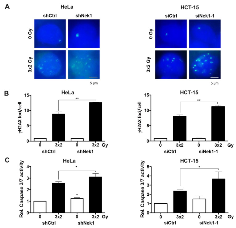
Figure 3.
(A) Representative images of γH2AX foci from non-irradiated and 3 × 2 Gy-irradiated control and Nek1 KD Hela and HCT-15 cells are shown. Nuclei were counterstained with DAPI. Scale bar, 5 μm. (B) Residual γH2AX foci and (C) apoptosis induction (24 h after the last fraction) of Dox-treated HeLa shCtrl and shNek1 (left) and HCT-15 cells (right) transfected with non-specific Ctrl and Nek1 siRNAs following a 3 × 2 Gy and 6 h interval fractionated irradiation (means ± SD; n = 3; * p < 0.05, ** p < 0.01 Nek1 KD vs. control).

