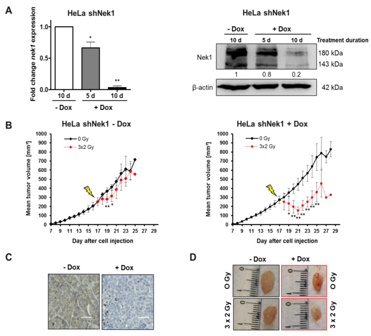
Figure 4.
Female, 12- to 16-week-old NOD/SCID (NSG) mice were injected subcutaneously with 1 × 10⁶ HeLa shNek1 and HeLa shCtrl cells in 100 μL PBS. After visual detection of tumor nodes about 10 days post cell injection, Dox was given (2 µg/mL doxycycline hyclate + 2% sucrose) in drinking water for an additional 10 days. Mice were irradiated by image-guided-radiotherapy (IGRT) with three fractionated single doses of 2 Gy every 6 h to reach a total dose of 6 Gy. (A) Tumor Nek1 mRNA and protein expression was analyzed by quantitative PCR (left) and Western blotting (right) after a 5 days or 10 days treatment with Dox. Numbers indicate protein expression relative to β-actin control and normalized to 10 d–Dox. (B) Relative tumor growth curves as monitored by using calipers (6 animals per group) inoculated with HeLa shNek1 cells treated with (right) or without Dox (left). Mean values of tumor volumes for each treatment group are shown (* p < 0.05, ** p < 0.01, irradiated vs. non-irradiated mice). (C) Representative images of histological detection of Nek1 levels in tumor tissue (bars correspond to 50 μm) and (D) representative images of tumors after fractionated irradiation with 3 × 2 Gy in Nek1-depleted (+Dox) and non-depleted tumors (-Dox) at day 28 after cell injection.

