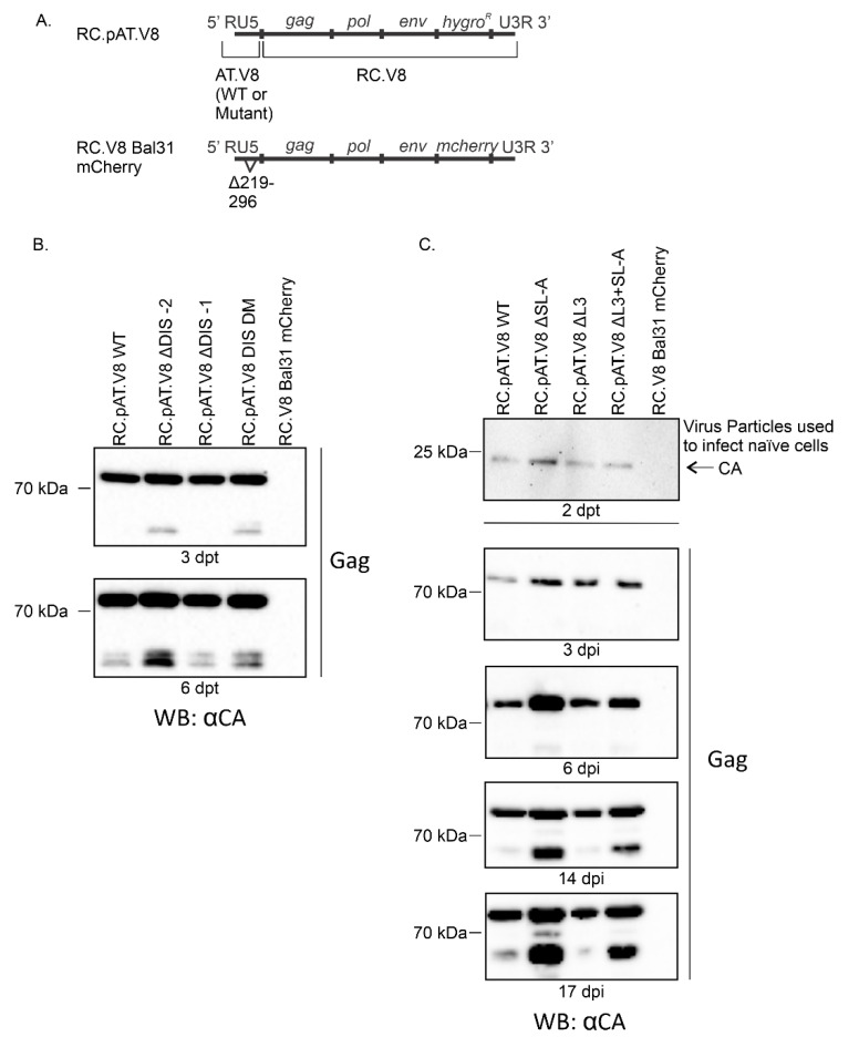
Figure 3.
(A) Schematic of the constructs used in the infectivity studies. (B) Immunoblots of Gag in 50 µg of lysates collected from QT6 cells transfected with RC.pAT.V8 WT, RC.pAT.V8 ∆DIS-2, RC.pAT.V8 ∆DIS-1, RC.pAT.V8 DIS-DM or RC.V8 Bal31 mCherry. Lysates were collected in RIPA at 3 and 6 dpt. (C) Immunoblots of virions collected from QT6 cells transfected with RC.pAT.V8 WT, RC.pAT.V8 ∆SL-A, RC.pAT.V8 ∆L3, RC.pAT.V8 ∆L3+SL-A and RC.V8 Bal31 mCherry (top panel, 2 dpt), and immunoblots of Gag in 50 µg of lysates collected from QT6 cells infected with viruses produced from the transfected cells (bottom panel, 3, 6, 14 and 17 dpi).

