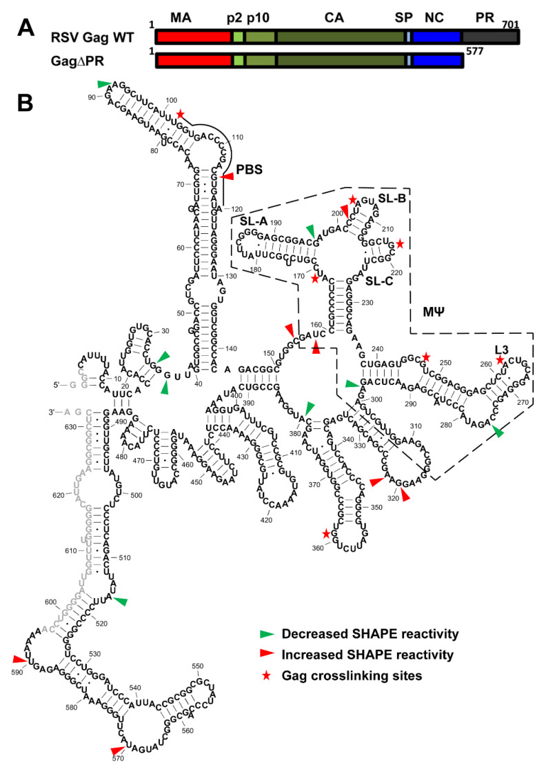
Figure 4.
Crosslinking (XL)-SHAPE analysis of RSV Gag∆PR binding to the RSV 5′-leader RNA. (A) Schematic showing individual domains of RSV Gag and RSV Gag∆PR. (B) XL-SHAPE results mapped to the secondary structure of the RSV 5′-leader RNA. Sites with decreased and increased SHAPE reactivity upon protein binding are indicated by green and red arrows, respectively. Identified crosslinking sites are labeled with stars. All identified sites have reactivity changes of ≥0.3 and p < 0.05 based on unpaired, two-tailed student t-tests, compared with the no protein control. The MΨ region (dashed box) and PBS are indicated. Results are based on the average of at least 3 independent experiments.

