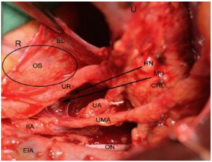
Figure 7.
Development of Okabayashi’s space during open surgery (right side of the pelvis). The ureter and mesoureter are separated from the posterior leaf of broad ligament. R—rectum; BL—posterior leaf of broad ligament; U—uterus; HN—right hypogastric nerve; OS—Okabayashi’s space development; MU—mesoureter; CRL—cardinal ligament; ON—obturator nerve; UMA—umbilical artery; UA—uterine artery; UR—ureter; EIA—external iliac artery; IIA—internal iliac artery.

