Figure 2.
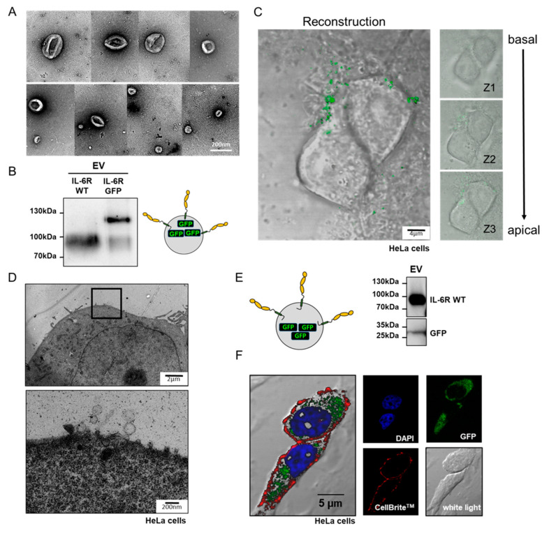
Isolated EVs interact with cells. (A) Transmission electron microscopic (TEM) image of EVs prepared from supernatants of transfected HEK293 ADAM10/17−/− cells. (B) Western blot analysis of EVs containing either untagged IL-6R (IL-6R WT) or IL-6R C-terminally tagged with green-fluorescence protein (IL-6R GFP). (C) Z-stacking reconstruction using confocal microscopy images of EVs containing IL-6R-GFP that were added to HeLa cells. (D) TEM image of an ultrathin cut HeLa cell incubated with EVs containing the IL-6R. EVs interacting with the cell surface could be observed at higher magnification. (E) Control Western blot of EVs carrying the IL-6R and a soluble version of GFP. (F) Confocal microscopy image of HeLa cells incubated with vesicles depicted in E. The cell nucleus is stained in blue (Bisbenzimide), the cell membrane is stained using CellBriteTM (red), GFP is in green, and a white light image was taken to display cell contours.

