Figure 3.
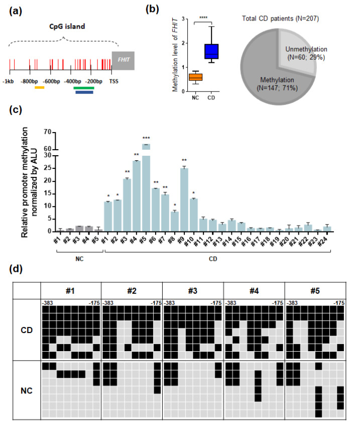
Summary of methylation analyses of the fragile histidine triad (FHIT) in CD tissues. (a) A schematic representation of the CpG islands in the FHIT gene promoter region. Each CpG site is indicated as a vertical red line, and the yellow, green, and blue boxes indicate the CpG probe site, amplicons for MSP, and bisulfite sequencing, respectively. (b) Methylation analysis of FHIT in patients with CD samples. Methylation level of FHIT between CD tissues and normal colon tissues (n = 12 each) and a Venn diagram indicates the methylation frequency of FHIT in a larger cohort of patients with CD (n = 207). (c) Quantitative MSP of the FHIT gene in selective ulcerative colitis (UC) patient samples and controls. All quantitative methylation levels were normalized by the Alu element. The statistical significance (p < 0.001) for the FHIT gene is shown between patients with CD samples. (d) Bisulfite sequencing analyses of the CpG islands in FHIT gene promoter regions. Bisulfite sequencing analyses were performed with representative patients with CD samples (CD; n = 5) and normal colon tissues (NC; n = 5). The location of CpG sites in the FHIT (upstream region from −383 to −175) relative to the transcription start sites (TSSs) of exon 1. Each box represents a CpG dinucleotide. Black boxes represent methylated cytosines and gray boxes represent unmethylated cytosines.

