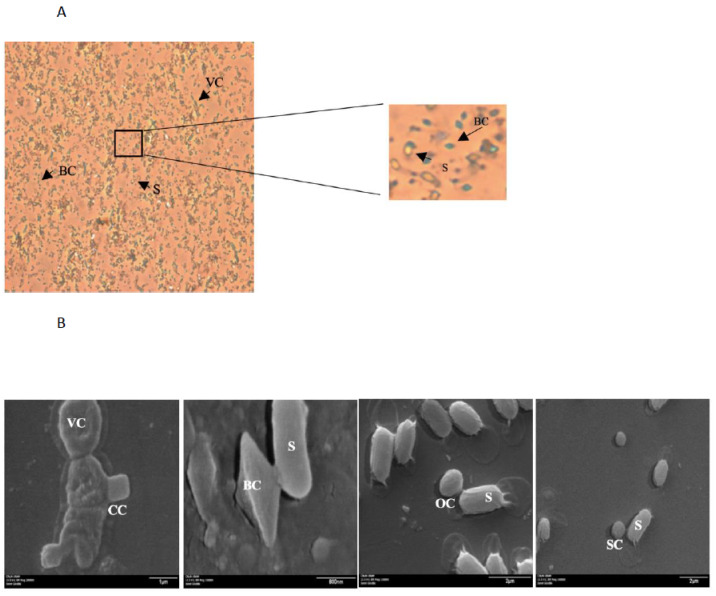
Figure 1.
Different morphologies of Bacillus thuringiensis crystals; (A) image observed at 40× in an optical microscope, the crystal was stained with malachite green. (B) Image observed at SEM with different magnifications from the left to the right (1) 20,000×, (2) 15,000×, (3) 15,000×, and (4) 50,000×. VC: vegetative cell; CC: cubic crystal; S: spore; BC: bipyramidal crystal; OC: ovoid crystal; SC: spherical crystal.

