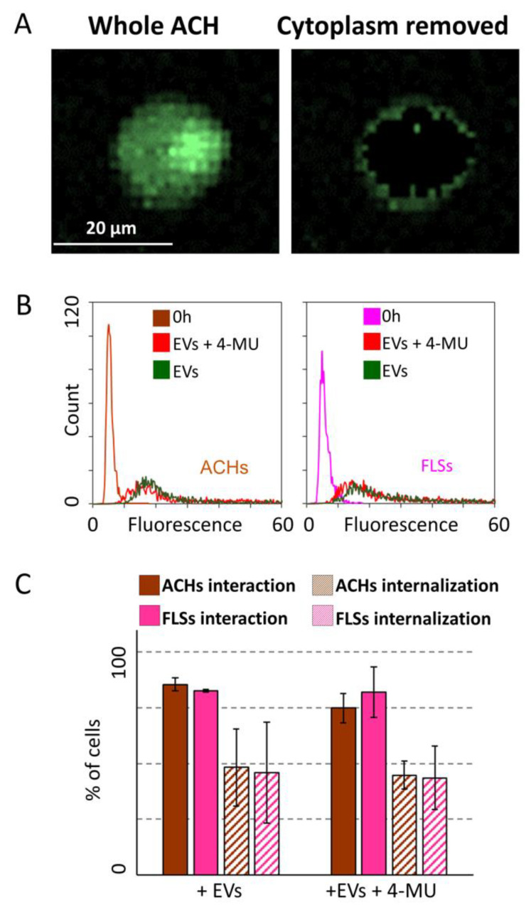
Figure 7.
HA removal effect on ASC-EV interaction with and incorporation in ACHs and FLSs in 3D microfluidic devices. (A) Image of a selected ACHs before and after removal of fluorescence signals associated with the cytoplasm. The presence of EVs outside the cell body is clear. (B) EV fluorescence intensity at time 0 and 24 h of ACHs and FLSs treated or not with 4-MU inside the 3D microfluidic device. 4-MU treated samples at time 0 are not shown for clarity, since no differences with control samples at time 0 emerged. N = 3 (C) ASC-EVs interacting with or internalized in ACHs or FLSs with or without treatment with 4-MU at 24 h. N = 3, values are shown as mean ± SE. Significance for p-value < 0.05.

