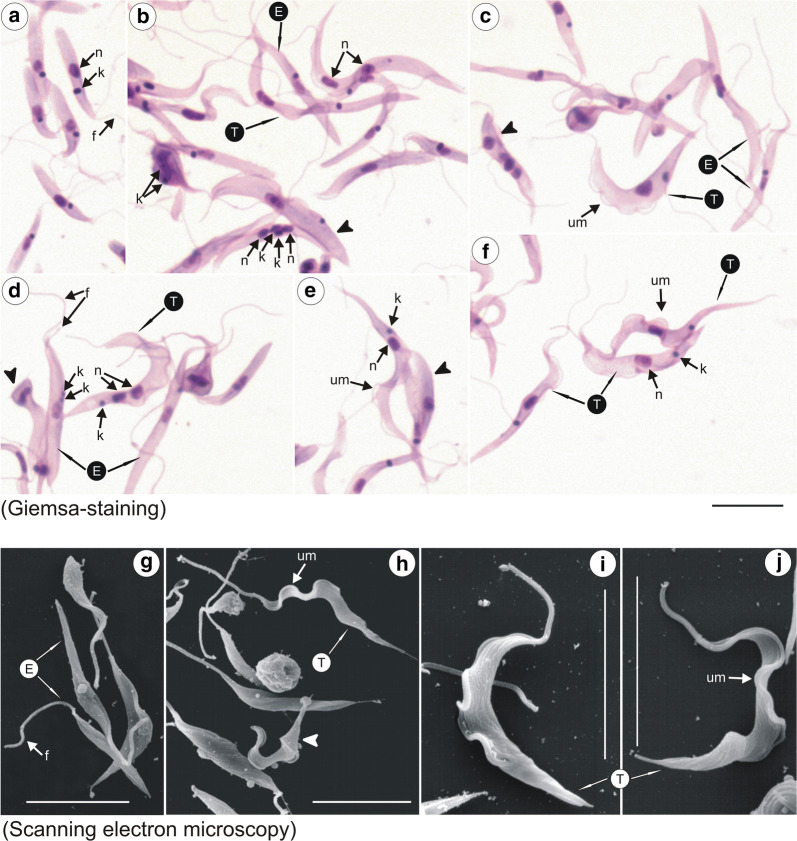
Fig. 8.
Light (Giemsa-staining) and scanning electron microscopy (SEM) of Trypanosoma trinaperronei n. sp. cultured with monolayers of mammalian (LLCMK2) cells at 37 °C. Giemsa-stained (a–f) and SEM (g–j) showing epimastigotes with one day of culture (a, g), and developmental forms (5 days): large and wide epimastigotes and trypomastigotes (b-d); flagellates with two nuclei and one kinetoplast (b, d); transition forms (arrow heads) between epimastigotes and trypomastigotes (b, d, e); and slender and pointed trypomastigotes with a well-developed undulating membrane (d–f, h–j).Abbreviations: nucleus, n; kinetoplast, k; flagellum, f; undulating membrane, um; epimastigote, E; trypomastigote, T; trypomastigote, T. Scale-bars: a–j,10 µm

