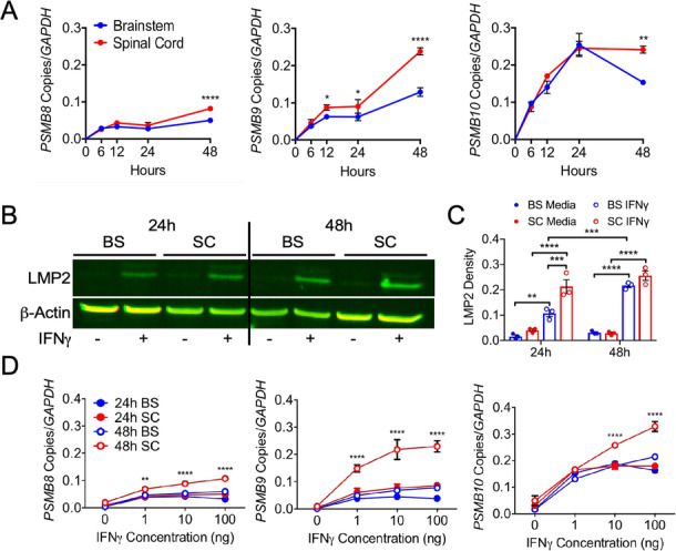
Fig. 2.

IFNγ-regulated expression of the iP in regional human astrocytes. a Human brainstem and spinal cord astrocytes were stimulated with 10 ng/ml IFNγ for 0, 6, 12, 24, or 48 h, and RNA was collected and analyzed for transcript levels of PSMB8, PSMB9, and PSMB10.b Human brainstem and spinal cord astrocytes were stimulated with or without 10 ng/ml IFNγ for 24 or 48 h, and protein lysate was assessed for levels of LMP2, normalizing to β-actin. c Human brainstem and spinal cord astrocytes were stimulated with 0, 1, 10, or 100 ng/ml IFNγ for 24 or 48 h, and RNA was collected and analyzed for transcript levels of PSMB8, PSMB9, and PSMB10. Data represent the mean ± SEM from 3 independent experiments. *P < 0.05, **P < 0.01, ***P < 0.001, ****P < 0.0001 by 2-way ANOVA. BS brainstem, SC spinal cord
