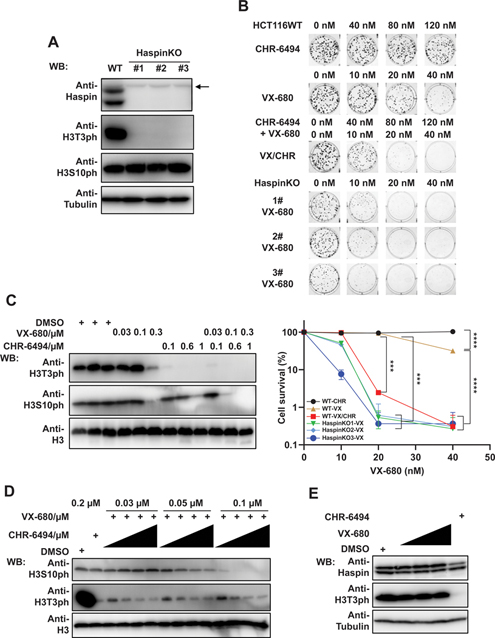
Figure 2.
Depletion or inhibition of Haspin sensitizes cells to VX-680 treatment. (A) Confirmation of Haspin depletion in Haspin KO cell lines. HCT116 wild-type and Haspin KO cells were collected and directly lysed by SDS loading buffer. The expression of Haspin, H3T3ph, and H3S10ph were detected with indicated antibodies. Anti-Tubulin and anti-H3 blots, respectively, were included as loading controls for Haspin, H3T3ph and H3S10ph blots. The arrow indicates nonspecific bands. (B) Cells treated with Haspin depletion or inhibition exhibit hypersensitivity to VX-680 treatment. HCT116 wild-type and Haspin KO cells were treated with CHR-6494 and VX-680 for consecutive 9 days, either in combination (VX combined with 0, 40, 80, and 120 nM CHR) or single-agent treatment (CHR; VX). Colonies were fixed, stained, and further analyzed by Image J. Experiments were performed in triplicate with duplicate biological replicates. Representative images and results are shown. Student t tests were performed to estimate differences between 2 groups. Error bar represents SE (n = 3); *** P < 0.001; **** P < 0.0001. (C) HCT116 cells were treated with indicated concentrations of VX-680 and CHR-6494 and further incubated for 24 h. Then cells were lysed by SDS loading buffer and examined by immunoblotting with indicated antibodies. The H3T3ph and H3S10ph levels were determined with indicated antibodies. H3 served as loading control. (D) HCT116 cells were incubated with VX-680 (0.03, 0.05, and 0.1 μM) and CHR-6494 (0.05, 0.1, 0.15, and 0.2 μM) in combination or single agent treatment. After 24 h, cells were lysed and examined by Western blotting with indicated antibodies. (E) Western blots show the effect of single-agent treatment (0.03, 0.05, and 0.1 μM VX; 0.2 μM CHR). Samples were collected and blotted as described in (D).

