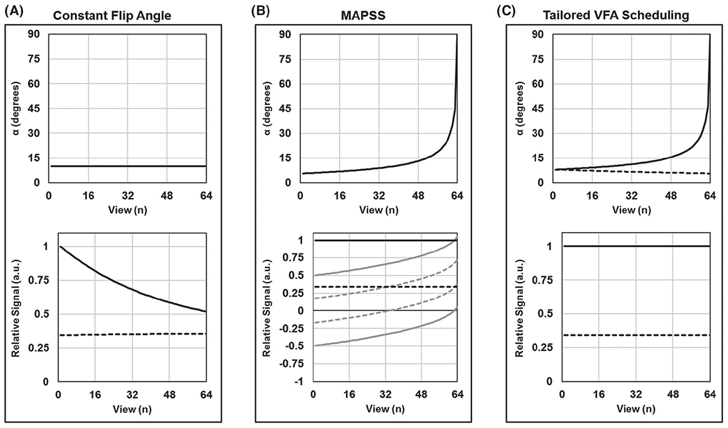
FIGURE 2.

Comparison of variable flip angle (VFA) techniques for segmented gradient-echo (GRE) T1ρ mapping. Results are simulated for VPS = 64 and TSL = 0 (solid lines) and 80 ms (dashed lines). Flip-angle schedules are shown in the top plots, and corresponding k-space signal modulations along the phase-encode dimension are shown in the bottom plots (each plot is individually normalized). A, If a constant flip angle is used to acquire all views following a spin-lock preparation pulse, then the measured signal will be modulated across the k-space phase-encoding plane. In this case (αn = 10°), TSL = 0 ms will produce a low-pass filtered image and TSL = 80 ms will produce a slightly high-pass filtered image. B, Magnetization-prepared angle-modulated partitioned k-space spoiled GRE snapshot (MAPSS) corrects for the k-space signal modulation by combining RF cycling with a single VFA train that is the same for all TSLs. Each TSL image is acquired twice, with magnetization, respectively, prepared along the +z and −z axes (gray lines); these signals are then subtracted to yield constant signal amplitude across the k-space phase-encoding plane (black lines). C, Tailored VFA scheduling corrects for the k-space signal modulation by applying a unique VFA train for each TSL, which obviates the need for RF cycling. VFA trains are generated based on assumed brain-tissue T1 and T1ρ relaxation times, such that each TSL image ideally has a constant signal amplitude across its k-space phase-encoding plane. Note that the VFA trains at TSL = 0 are different for MAPSS and tailored VFA scheduling, with different α1
