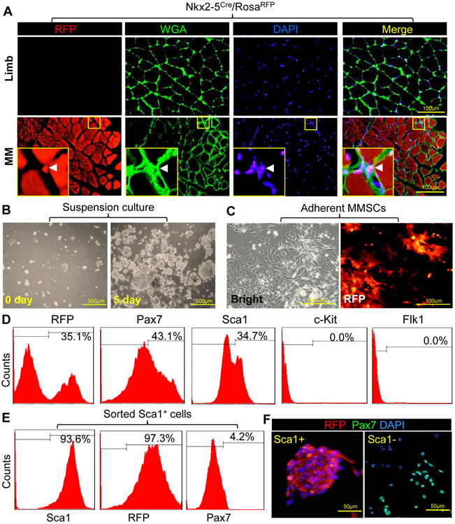
Figure 1. Identification and characterization of MMSCs.
(A): Limb or masseter muscles (MM) were isolated from Nkx2-5Cre/RosaRFP mice. Tissue sections stained with wheat germ agglutinin (WGA-FITC, green) were observed under multiple channels on a confocal microscope. Arrow head in magnification view indicates the location of RFP+ MMSCs. (B): MMSCs were isolated and maintained in suspension culture. (C): The expression of RFP was observed by microscope after culture of MMSCs in monolayer. (D): Surface markers and RFP in MMSCs were analyzed by FACS after primary cell isolation. Flow cytometric gates (indicated by solid lines) were set using the appropriate isotype control antibody. (E): RFP and Pax7 expressions in Sca1+ MMSCs were analyzed by FACS after sorting. (F): Immunostaining of Pax7 and RFP expression in sorted cells was observed by microscope. Nuclei were stained with DAPI.

