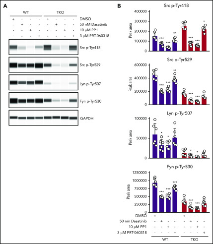
Figure 6.
SFKs auto-phosphorylate their C-terminal inhibitory tyrosine residues. (A) Representative blots of capillary-based immunoassays on platelet lysates with the indicated antibodies. Lysates were generated from platelets incubated with DMSO, 50 nM dasatinib, 10 μM PP1, or 3 μM PRT-060318 for 15 minutes, room temperature. (B) Quantification of peak areas, n = 6 mice/genotype. Asterisks refer to significant difference compared with DMSO-treated control samples within genotypes. *P < .05, **P < .01, ***P < .001, 2-way ANOVA with Sidak test; mean ± SD.

