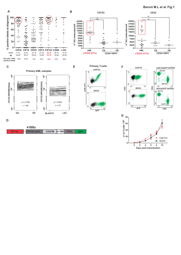
Figure 1.
Expression of CD123 in AML and design, detection and expansion CD123 CARTs. (A) Immunophenotyping of the indicated diagnostic myeloid markers in a cohort of 97 patients with AML at presentation. Each dot denotes an individual patient. Red circles identify patients with >80% of blasts positive for the indicated marker. (B) Comparative antigen density (measured as antigen molecules/cell) for CD123 and CD33 in primary AML samples (n=24), CB-derived (n=22) and PB-derived (n=10) CD34+ cells from healthy donors. AML blasts were identified as 7AAD−CD3−CD45+/lowCD123+CD33+. One-way analysis of variance; *p<0.05, **p<0.01, ***p<0.001. (C) Comparison of CD123 expression in 68 paired diagnostic-relapse AML samples (left panel) and in bulk tumor versus AML-LSC (n=37, right panel).33 (D) Scheme of the CD123 CAR structure. (E) CAR detection in primary T-cells using an antihuman IgG F(ab')2 antibody and GFP. (F) Successful CAR123 transduction and detection in CD4+ and CD8+ T-cells (n=3). (G) Robust expansion of activated T-cells transduced with either MOCK (black line) or CAR123 (red line) (n=3). AML, acute myeloid leukemia; CAR, chimeric antigen receptor; CART, chimeric antigen receptor T-cell; CB, cord blood; DX, diagnostic; GFP, green fluorescence protein; LSC, leukemia stem cell; PB, peripheral blood; RX, relapse.

