
Figure 1.
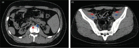
(a) CT image shows mirror‐image location of the abdominal organs and bilateral atrophic kidneys. Ventral aorta (red arrow) and inferior vena cava (blue arrow) are reversed left and right. (b) The external iliac arteries (red arrows) and veins (blue arrows) are shown. Right side vessels are deeper than left side ones (blue dashed line) (Case 3).