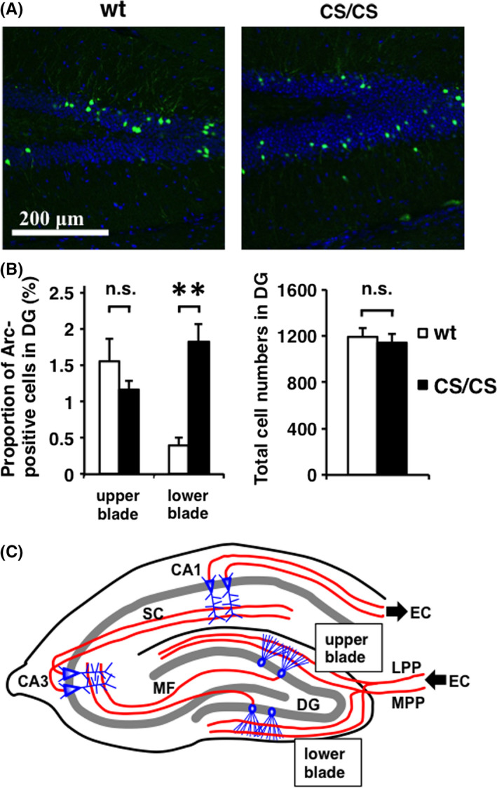
Figure 2.

Different Arc expression in the upper and lower blades of the hippocampal dentate gyrus at basal level. (A) Arc expression (green) and DAPI staining (blue) in wild‐type (wt) and GluA1C811S homozygous (CS/CS) mice. Typical patterns are shown. (B) Proportions of Arc‐positive cells in the upper and lower blades (left) and total cell numbers (DAPI‐positive cells, right) in the dentate gyrus (DG) of wt and CS/CS mice. Wt: n = 4, CS/CS: n = 4. Error bars represent SEM **P < 0.01, t test. n.s.: not significant. (C) Schematics of major excitatory circuits in the hippocampus. The hippocampal circuits consist of a trisynaptic pathway. Granule neurons in the dentate gyrus (DG) receive excitatory input from the entorhinal cortex (EC) via the medial perforant pathway (MPP) and lateral perforant pathway (LPP). Then, the granule neuron sends the signal to CA3 pyramidal neurons via the mossy fibers (MF). CA3 pyramidal cells relay the signal to CA1 pyramidal cells via Schaffer collaterals (SC). Finally, CA1 pyramidal neurons return the signal to the EC. See also Table S2
