. 2020 Jun 12;128(6):067010. doi: 10.1289/EHP6508

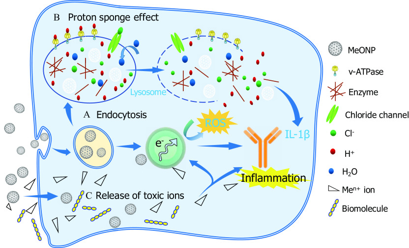
Figure 8.

Figure 8 is a schematic diagram representing image of inflammatory mechanisms by metal oxide nanomaterials in three stages. Stage a titled Endocytosis includes MeONPs, leading to electrons and ross, leading to IL–1 beta. Stage b titled Proton sponge effect includes lysosomes, comprising v-ATPase, MeONPs, enzymes, positively charged H ions, negatively charged Cl ions, H subscript 2 O, and chloride channel, leading to each other. Stage c titled release of toxic ions includes biomolecules and Me superscript n positive ions, leading to electrons and IL-1 beta and inflammation in stage a. Stage a leads to stage b and stage b leads back to stage a. MeONPs are the input to stages a and c.

Proposed schematic image of inflammatory mechanisms by metal oxide nanomaterials (MeONPs). (A) Endocytosis: MeONPs with a positive ζ-potential were most internalized by THP-1 cells and lysosomes. (B) Proton sponge effect. MeONPs with metal atom electronegativity 1.55 tend to trigger a proton sponge effect, followed by lysosome damages, leakage of lysosomal contents and excess IL-1β production. (C) Release of toxic ions.