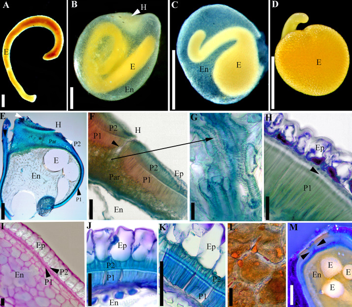
Fig 3. Seed features viewed with light microscopy.
A–D. Embryos. A. Cuscuta monogyna (embryo removed from the endosperm); note that in this species the embryo does not form a full coil. B. Embryo of C. tinctoria var. floribunda embedded in the endosperm. C. Developing embryo of C. nevadensis surrounded by the endosperm epidermis (the rest of endosperm was almost entirely consumed). D. Fully developed embryo of C. nevadensis (endosperm epidermis removed). E–H. Cuscuta lupuliformis (subg. Monogynella). E–G. Longitudinal sections through the hilum area of C. lupuliformis. E. Overview; black asterisk indicates position of water gap. F. Detail of hilum area; black arrow indicates position of water gap; note that two palisade layers (P1 and P2) are present. G. Tracheid-like structures embedded in a parenchyma tissue. H. Testa architecture outside hilum area with only one palisade layer; black arrow indicates linea lucida. I–K. Seed coat architecture with two palisade layers outside the hilum area. I. Incipient stage in the development of the two palisade layers in C. argentinana; at this stage, epidermis contains starch grains. J. Cuscuta europaea. K. Cuscuta cristata; note the presence of linea lucida in the inner palisade layer (P1). L. Parenchyma cells with lipids and starch in the enlarged portion of C. nevadensis embryo. M. Longitudinal section of rehydrated C. sandwichiana seed after 15 min in Aniline Blue; dye penetration is limited to the water gap (indicated with arrows). E = Embryo; En = endosperm; H = hilum; Ep = epidermis; Par = parenchyma; P1 = Inner or single palisade layer; P2 = Outer palisade layer. Scale bars. A–E = 0.5 mm; G, I–K = 50 μm; H, L = 25 μm; F, M = 100 μm.

