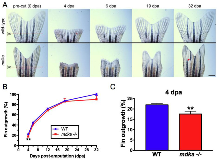
Fig 2. Initial fin regeneration is delayed but recovers in mdkami5001.
(A) Fins from wildtype (top) and mdkami5001 (bottom) at 0, 4, 6, 19, and 32 dpa. Red lines indicate the level of the amputation plane. In wildtype animals, the structure of the bony fin rays and pigmentation recover completely. Compared to age-matched group of wildtype, pre-amputated fin in the mdkami5001 is smaller in size (wildtype: 51.3 ± 7.1 μm2; mdkami5001: 34.3 ± 4.7 μm2, p<0.0001, Student’s t-test). In the mutants, regenerated fins display abnormal shape and mis-patterned melanophore pigmentation (red arrow). Scale bar equals 400 μm. (B) Graph illustrating the average percent outgrowth through 32 dpa, calculated from the ratio of the areas of pre- and post-amputation fins. Wildtype fins recover to 100% of pre-amputation levels by 32 dpa. The mutant animals show a significant reduction of fin outgrowth at 4 dpa. At the subsequent time points, there is no statistically significant difference in the fin outgrowth between wildtype and mdkami5001. (C) Graph illustrating the percent fin outgrowth at 4 dpa in wildtype and mdkami5001. Student’s t-test, p = 0.0105. n = 9 wildtype and 8 mutants.

