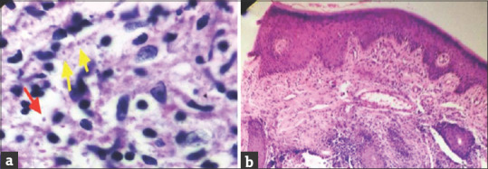
Figure 3.

(a) Slit-skin smear with Leishmania donovani (LD) bodies in the intracellular (yellow arrows) and extracellular spaces (red arrow) ( Giemsa, ×1000). (b) Skin biopsy showing dense inflammation in the dermis along with noncaseating epitheloid granuloma (black arrows) in the dermis (H and E, ×100)
