Fig. 5.

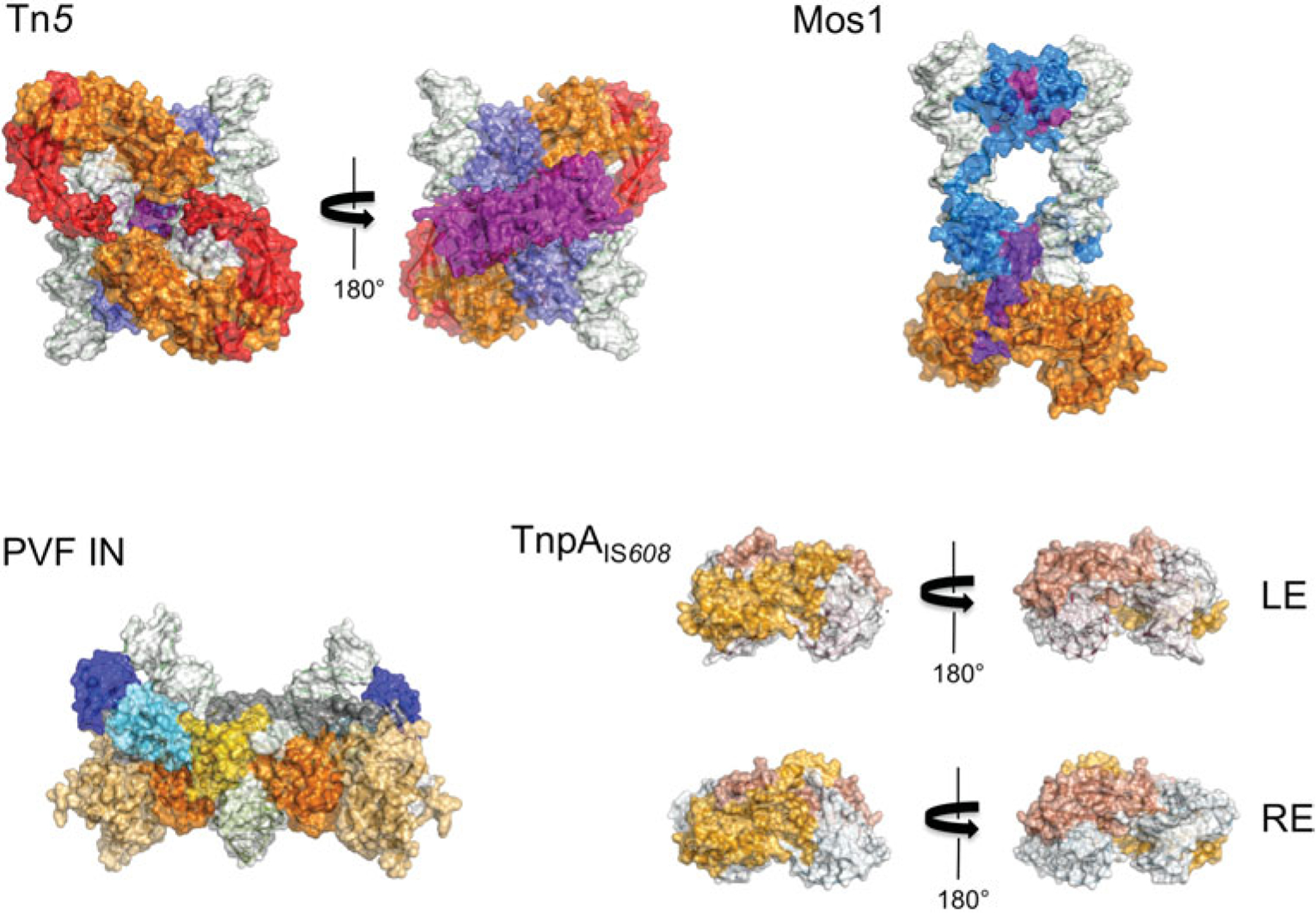
Space-filling representation of transpososomes (to scale). The domain colors correspond to those in Fig. 2. DNA is shown with a white surface. In the case of PVF integrase, a target capture complex is shown.

Space-filling representation of transpososomes (to scale). The domain colors correspond to those in Fig. 2. DNA is shown with a white surface. In the case of PVF integrase, a target capture complex is shown.