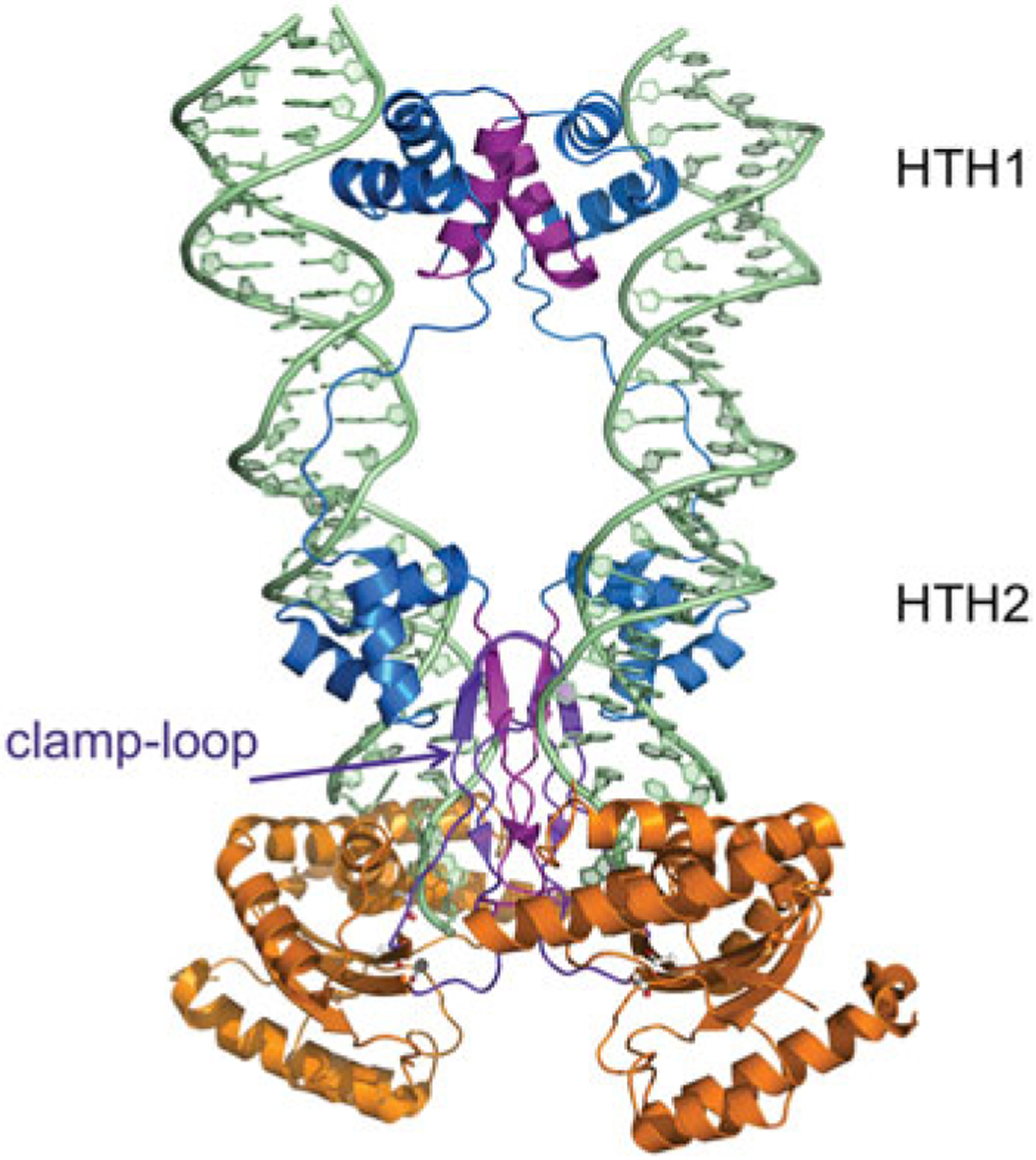
Fig. 7.

The dimeric Mos1 transpososome. The domain colors correspond to those shown in Fig. 2. The N-terminal DNA domain (two HTH domains connected by a long linker) is in blue and the RNaseH-like catalytic core is in orange. Residues involved in multimerization (amino acids 7–21, 112–125, and the clamp–loop residues 162–189) are shown in purple. The residues comprising the DDD motif (D156, D284, and D249) are shown in ball-and-stick representation. PDB code: 3HOT.
