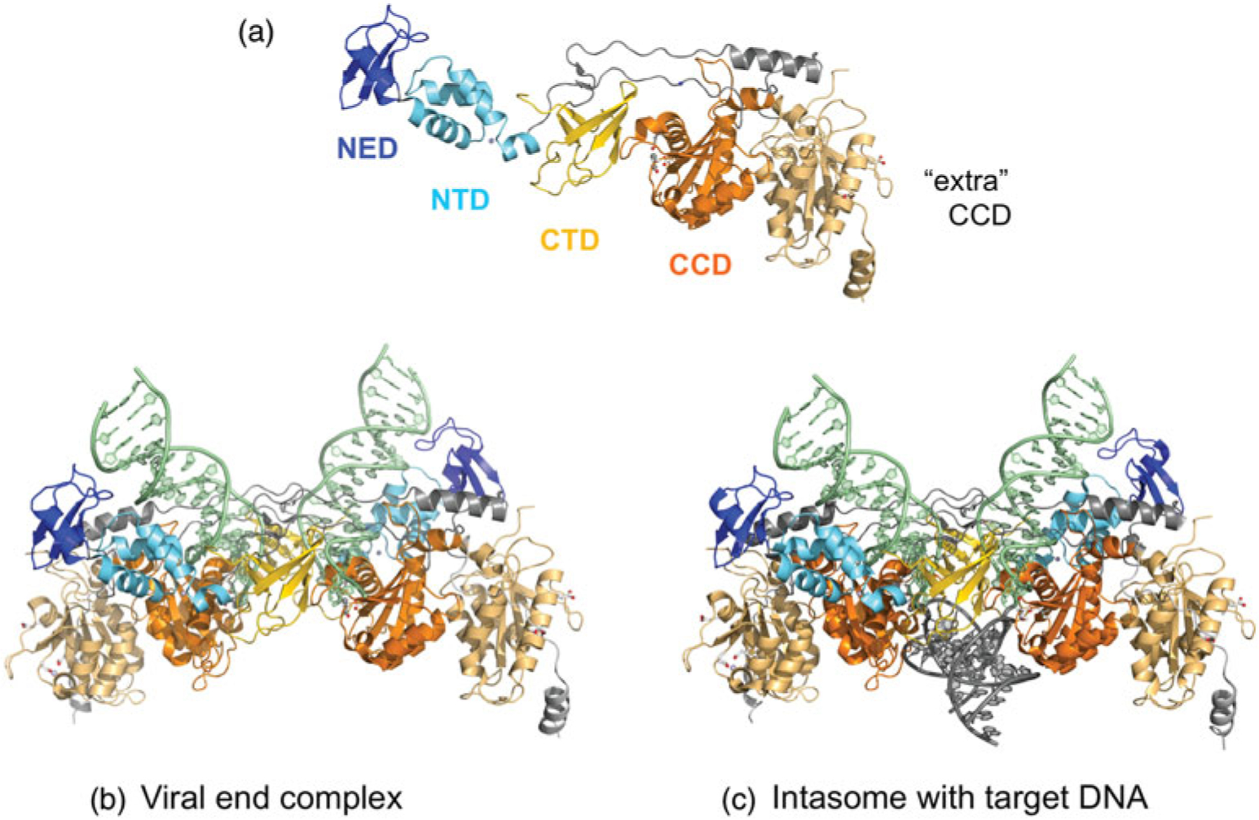
Fig. 8.

The PFV intasome. (a) The extended string of domains of one monomer of the intasome (domains in color) and its associated unused or ‘extra’ catalytic domain (in light orange). Below are the structures of (b) the intasome with two viral DNA ends and (c) the STC. The domain colors correspond to those shown in Fig. 2, where dark blue is the NED, light blue is the Zn-binding NTD (the Zn2+ ion is shown as a light blue sphere), orange is the CCD (there are two Mn2+ ions bound in the ‘used’ active site), and yellow is the CTD. The two additional CCDs with unused active sites on the periphery of the intasome contributed by outer subunits are shown in light orange in (b) and (c). PDB codes used to generate intasome figure with viral ends (3OY9) and with target DNA (3OS0).
