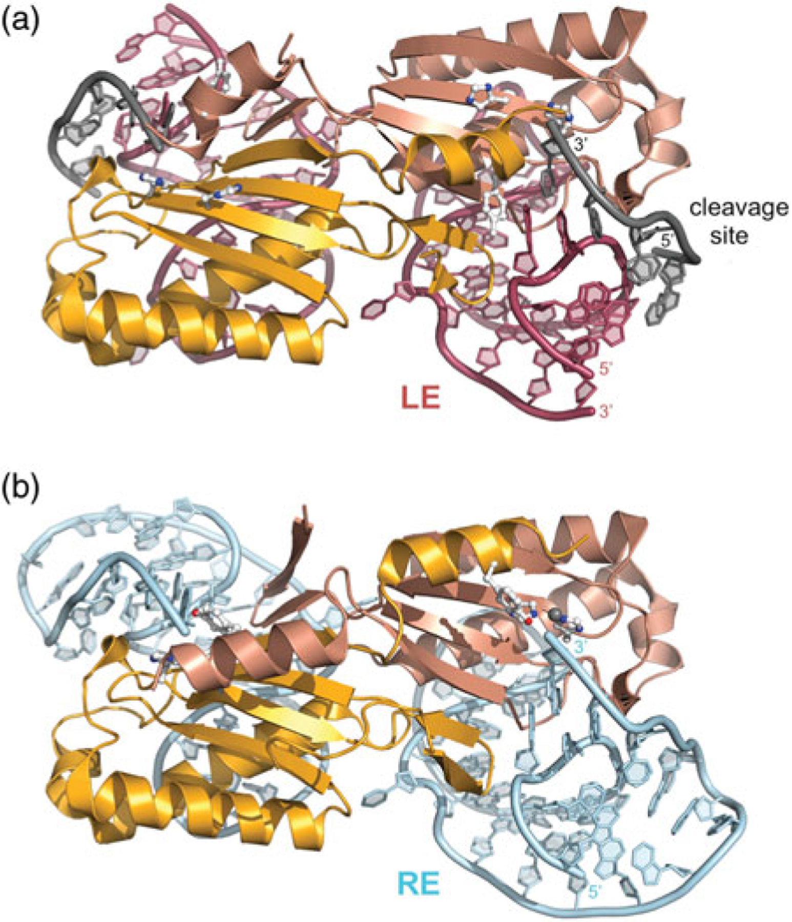
Fig. 9.

Dimeric IS608 TnpA transpososomes. (a) TnpA bound to an LE 26-mer (in red) and a 6-mer cleavage or target site (grey). The two monomers are shown in different shades of orange to illustrate the composite active site. Active site residues H64, H66, and Y127 are shown in ball-and-stick representation. PDB code: 2VJV. In this structure, Y127 has been replaced by Phe. (b) TnpA bound to a RE 35-mer (in blue). PDB code: 2VJU.
