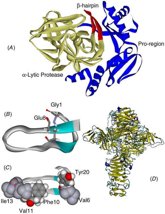
Figure 2.
(A) The complex of α-lytic protease (yellow ribbon) with its proregion (blue ribbon); the β-hairpin (red ribbon) enhances the kinetic stability of α-lytic protease (PDB code: 4pro). (B) Two chemically linked residues in microcin J25 (PDB code: 1pp5). (C) Hydrophobic surface in microcin J25. (D) The dominating β-sheet and β-barrel structure of the endosialidase trimer (PDB code: 1v0e).

