
Fig. 6. Infiltration of lymphocytes in SLE kidneys.
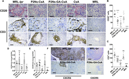
(A) Histological images showing that renal damage in the untreated MRL-lpr appeared to have a strong correlation with lymphocyte infiltration. Both CD20+ and CD3+ cells were enriched in the glomeruli (CD20 and CD3) and the interstitial space (CD3) (n = 15 to 30 micrographs from at least three individuals; scale bars, 50 μm for first two rows of images; scale bars, 200 μm for third row of images). (B) Graphical analyses of CD20+ cells and (C) CD3+ cells in the histological images. (D) We observed elevated transcript levels of CXCR4 and (E) CXCR5 in the untreated MRL-lpr mice (n = 5 to 6 individuals). (F) Renal histology also showed significant up-regulation of CXCR4 (brown) among infiltrating leukocytes and endogenous expression in the proximal tubules. CXCR5+ population (red) appeared to decorate the migrating front of infiltrating leukocytes. (scale bars, 200 μm for all images). Representative micrographs are shown. Data are represented as mean ± SEM. *P < 0.05, **P < 0.01, ***P < 0.001, and ****P < 0.0001. Statistics were conducted against the MRL-lpr group. Comparisons were made with one-way ANOVA, followed by Tukey’s multiple comparisons test.