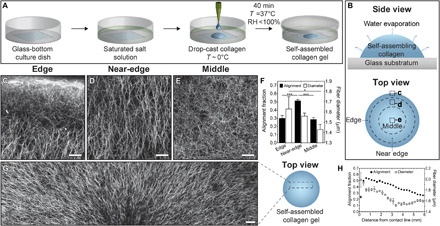
Fig. 1. Self-assembly of collagen in evaporating droplets generates aligned networks of collagen fibers.

Schematic of (A) drop-casting procedure and (B) top and side views of an evaporating droplet of collagen. CRM images of self-assembled droplet of collagen in the (C) edge, (D) near-edge, and (E) middle regions of interest. Images are oriented such that the top of the image points toward the contact line of the droplet. The location for each image is highlighted in dashed boxes in (B). Scale bars represent 50 μm. (F) Alignment fraction and fiber diameter for drop-cast collagen gels. (G) CRM image of a self-assembled droplet of collagen. Five separate CRM images are stitched together to reveal the radial alignment of collagen fibers. Scale bar, 100 μm. (H) Alignment fraction and fiber diameter as a function of distance from the contact line for drop-cast collagen gels. Collagen solutions (pH 11) were gelled at controlled RH using a saturated solution of MgCl2 (RH ~ 31%) on UVO-treated glass. *P ≤ 0.05 and ***P ≤ 0.001.
