. 2020 May 27;9:e57887. doi: 10.7554/eLife.57887

Figure 3. Functional analysis of the EMC2•EMC9 cytosolic vestibule.

(A) 35S-methionine-labeled TMD of SQS was mixed with recombinant purified EMC2•EMC9 complex as in Figure 1 and analyzed directly or after UV irradiation as indicated. The photocrosslinking amino acid benzoyl-phenylalanine (Bpa) was incorporated into either the SQS substrate, EMC9 (at codon 167), or EMC2 (at codon 193) as indicated. Both R167 in EMC9 and Q193 in EMC2 line the vestibule. (B) Bpa was incorporated at different positions within the vestibule of the EMC2•EMC9 complex and crosslinking efficiency to SQS was determined as in panel A. Locations of the Bpa are annotated as spheres on the EMC2•EMC9 heterodimer. The sphere colors correspond to the intensity of the resulting crosslink. Position 191 (obscured behind L190 in this view) showed no crosslinking to substrate, consistent with its rearward facing location. (C) Shown on the left is a diagram of the dual color reporter for insertion of the TMD of SQS. Expression of this reporter results in a free GFP protein and an RFP-tagged SQS protein due to ribosomal skipping at the a viral 2A sequence. The left graph shows flow cytometry analysis of the SQS reporter in WT cells (grey), EMC2 knockout (KO) cells (shaded pink), KO cells complemented with WT EMC2 (blue line), and KO cells complemented with the HLY* EMC2 mutant (red line). The data are represented as histograms of the RFP to GFP ratio. The right graph shows a comparison of the SQS reporter in KO cells complemented with either WT EMC2 or the IMA* EMC2 mutant. The mutated amino acids, whose positions are shown in panel B, are: H189E, L190E, Y191K, I61K, M95K, and A122E.

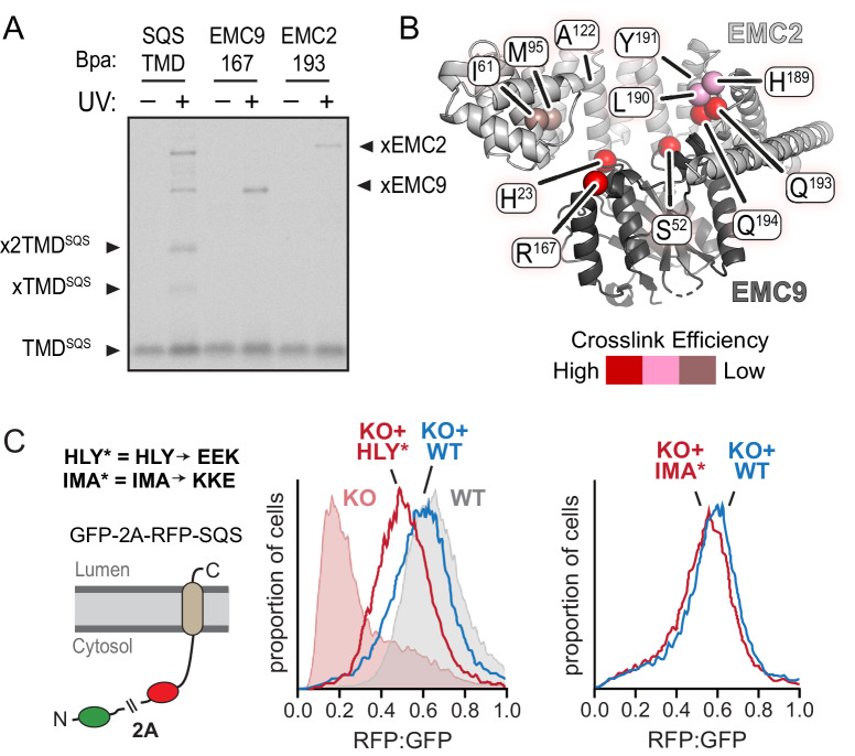
Figure 3.

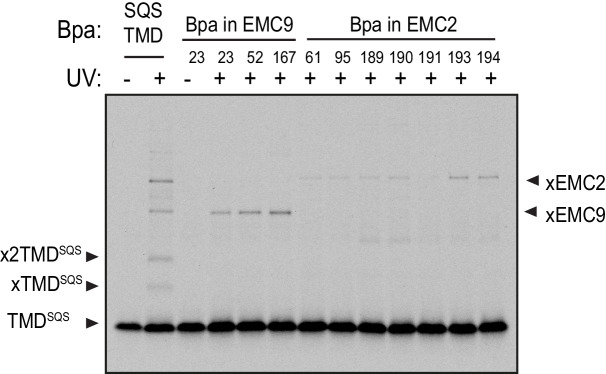
Figure 3—figure supplement 1. Crosslinking analysis of the EMC2•EMC9 cytosolic vestibule.

Figure 3—figure supplement 1.

35S-methionine-labeled TMD of SQS was mixed with recombinant purified EMC2•EMC9 complex as in Figure 1 and analyzed directly or after UV irradiation as indicated. The photocrosslinking amino acid benzoyl-phenylalanine (Bpa) was incorporated into either the SQS substrate, EMC9, or EMC2 as indicated.
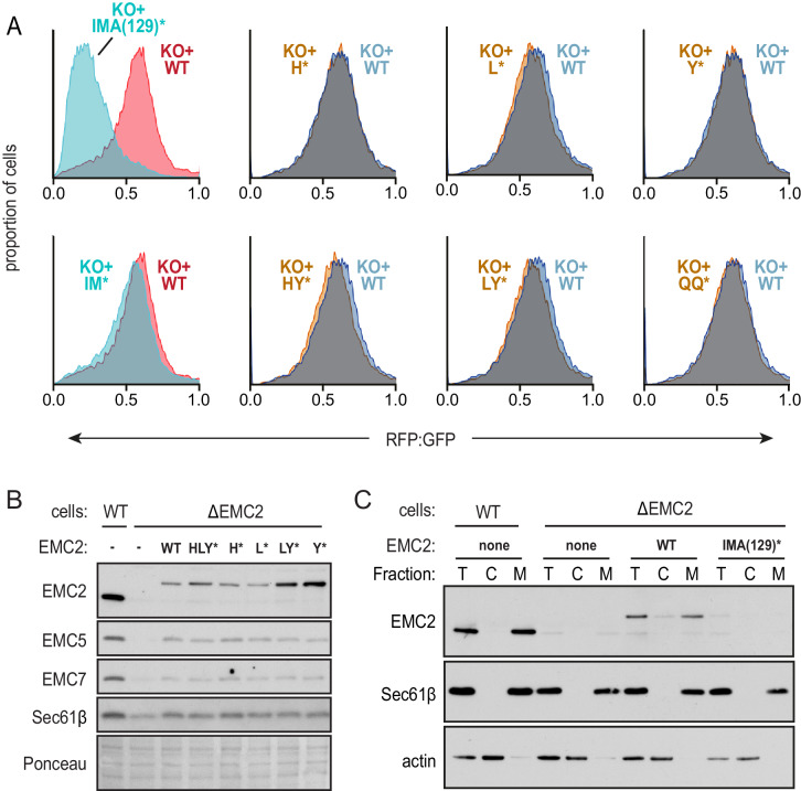
Figure 3—figure supplement 2. Functional analysis of the EMC2•EMC9 cytosolic vestibule.

Figure 3—figure supplement 2.

(A) Expression of the dual color SQS reporter in EMC2 knockout cells complemented with either WT EMC2 or the indicated EMC2 mutants. The data are represented as histograms of the RFP to GFP ratio. The two left graphs are from a different experiment from the remainder of graphs (which were analyzed together) and are colored differently to indicate this. The knockout-like phenotype of the IMA(129)* mutation is apparently due to the A129K mutation since the IM* mutation shows almost complete rescue. The mutated amino acids are: I61K, M95K, A129K, H189E, L190E, Y191K, Q193E, and Q194K. (B) Immunoblotting of whole cell extracts from WT or ΔEMC2 cells transfected with the indicated EMC2 constructs. A section of the Ponceau-stained blot shows equal loading. (C) WT and ∆EMC2 cells transfected with the indicated EMC2 constructs were fractionated into cytosol (C) and membrane (M) fractions and analyzed by immunoblotting for EMC2 relative to an equivalent amount of total (T) cell lysate. The membrane marker Sec61β and cytosolic marker actin were included as controls. Note that relative to WT EMC2 expressed in ∆EMC2 cells or endogenous EMC2 in WT cells, the IMA(129)* mutant is very poorly expressed. This is presumably due to its degradation secondary to inefficient assembly with other EMC subunits, explaining its strong phenotype in the flow cytometry assay.