
Figure 2. Abnormal muscle histology and absence of collagen VI staining in Case 1.
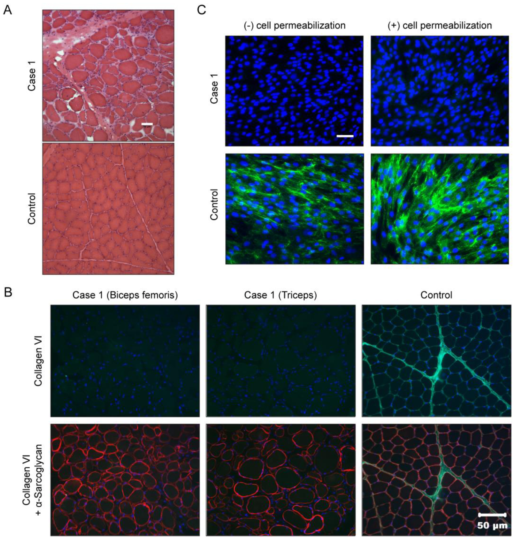
(A) Hematoxylin and eosin (H&E) staining of cryosections from the biceps femoris muscle from Case 1 showing a dystrophic phenotype including excessive variability in myofiber size, endomysial fibrosis and excessive internal nuclei. Scale bar = 50 μm. (B) Muscle biopsy sections co-stained for collagen VI (green) and α-sarcoglycan (red). Scale bar = 50 μm. (C) Primary dermal fibroblasts cultured and stained for collagen VI (green), and nuclei (DAPI, blue), in absence (−) or presence (+) of a cell permeabilizing agent. Scale bar = 50 μm.