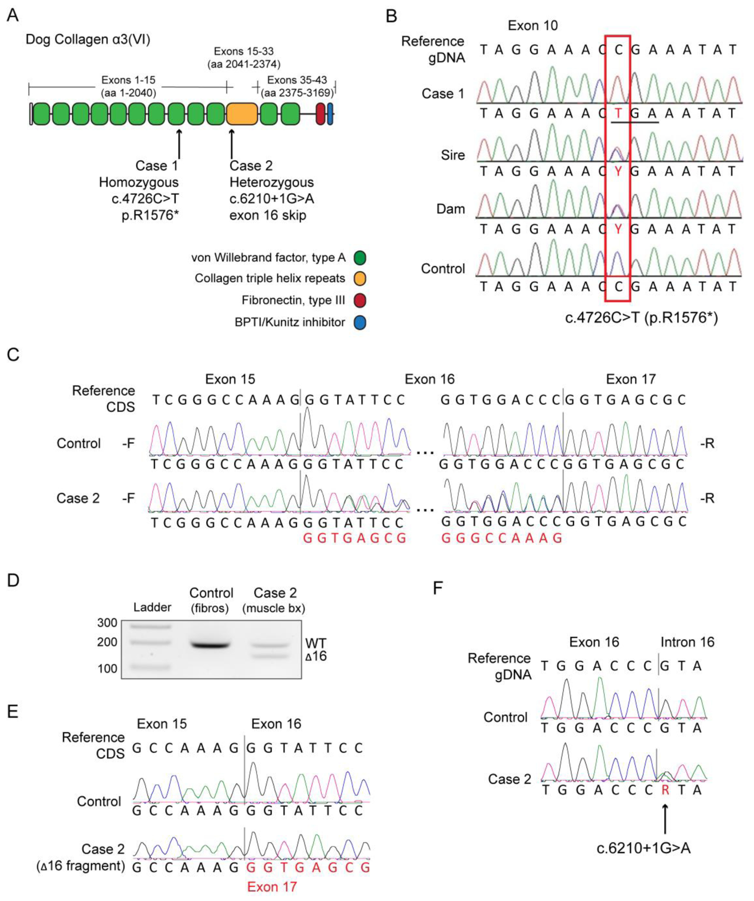
Figure 3. Muscular dystrophy in Labrador retriever dogs caused by variants in COL6A3.

(A) Schematic of the main collagen a3(VI) chain protein domains in the dog, and the relative positions of the recessive and dominant variants found in the affected Labrador retriever dogs Case 1 and Case 2, respectively. aa = amino acid. (B) Alignments of exon 10 sequences of Case 1, the sire, the dam, and a control obtained by Sanger sequencing of a PCR amplicon. The box indicates the location where a homozygous C nucleotide in the control genome is replaced with a homozygous T nucleotide in the case. Both the sire and dam are heterozygous. The variant changes the codon for an arginine residue (CGA) to TCA to the stop codon (TGA). (C) Complementary DNA (cDNA) forward (-F) and reverse (-R) sequencing chromatograms of a control dog and of Case 2, spanning exon 16 of the COL6A3 gene. CDS = coding sequence. (D) cDNA amplification and electrophoresis of COL6A3 transcripts. The fragments in Case 2 lane show an approximately equal expression of normal transcripts (wild-type, WT) and transcripts lacking exon 16 (Δ16), which is 54 nucleotides in size. (E) Sequencing chromatograms of the PCR fragments isolated from the electrophoresis gel in (B) showing the junction of exon 15 to exon 17. (F) Sequencing chromatograms from genomic DNA show that Case 2 is a heterozygous carrier of the COL6A3 c.6210+1G>A variant.
