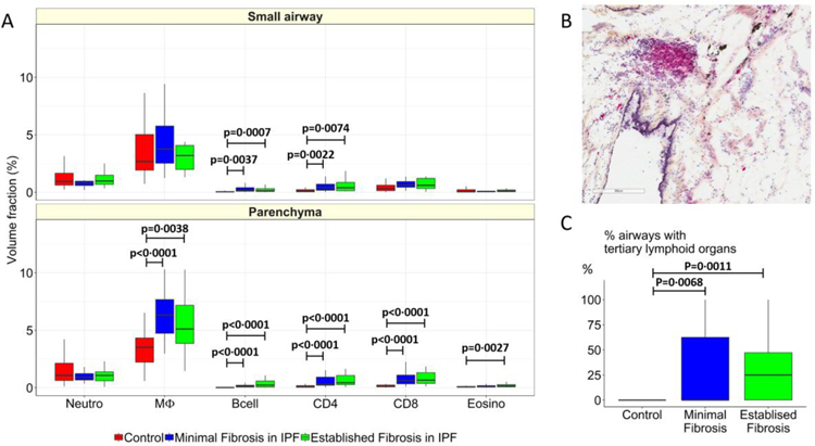
Figure 5:
Compares the volume fractions of infiltrating inflammatory immune cells in control lung tissue (red) to the volume fractions of the same cells in regions of minimal (blue) and established (green) fibrosis in experiment 2. This comparison shows (A) B-cell and CD4 lymphocyte infiltration increased in airways tissue and that the infiltration of macrophages, B-cells, CD4, CD8 and eosinophils are increased in the parenchyma in regions of minimal and established fibrosis compared to controls. These data show that the number airways containing tertiary lymphoid organs, as illustrated in (B), increased in both minimal and established regions of fibrosis compared to controls (C), but were not different from each other in the airways. Values are expressed as per sample.

