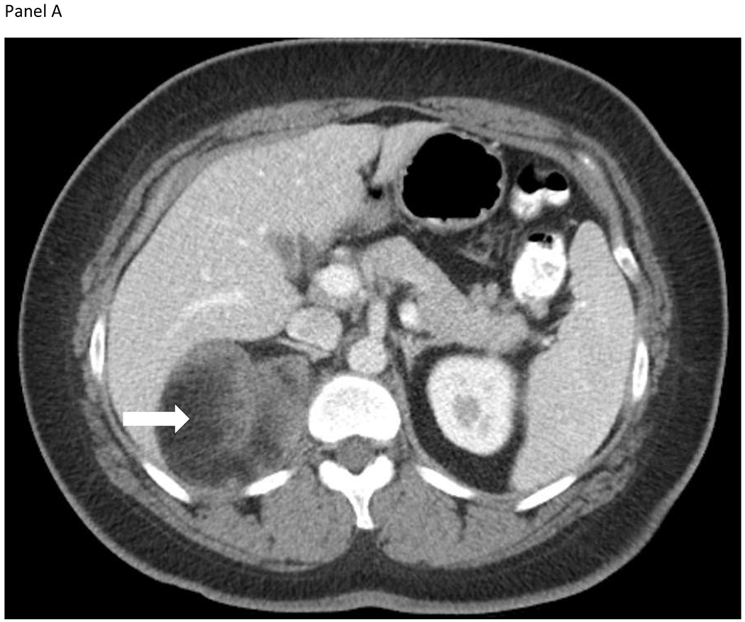
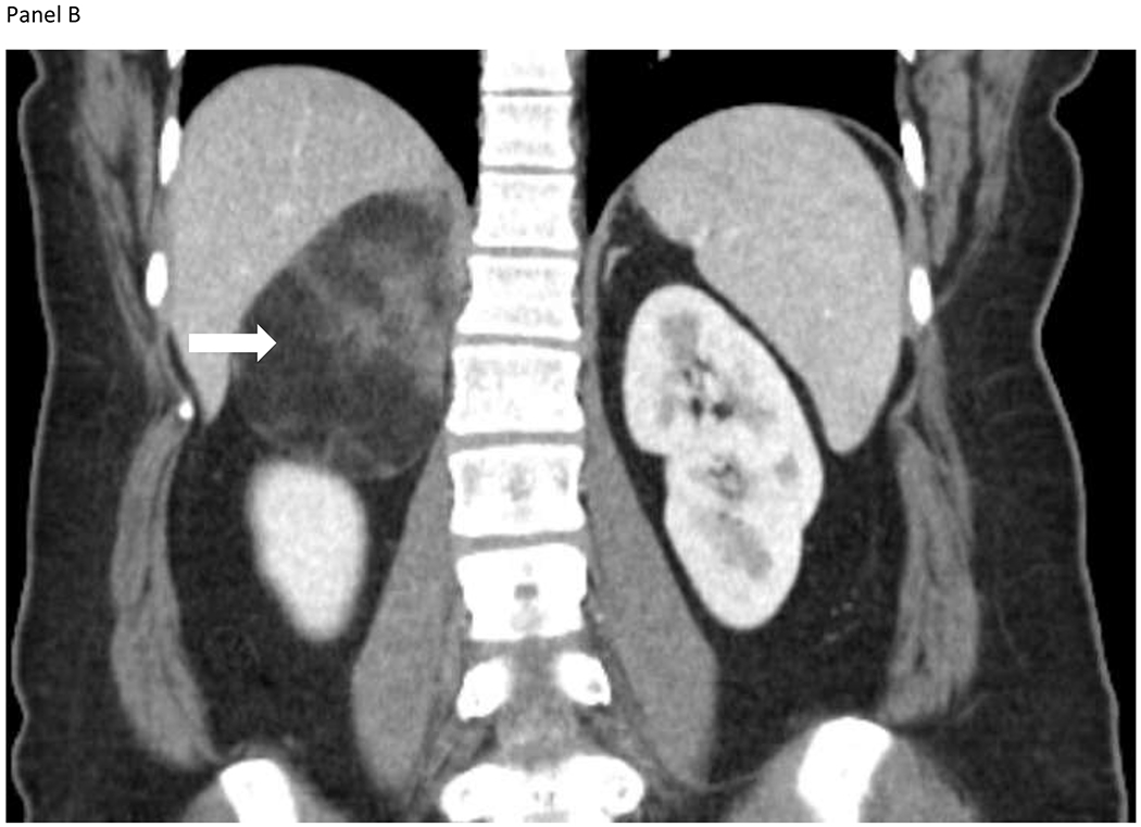
Figure 1.


A 33-year-old woman presented with intermittent right upper quadrant pain. She also reported constipation, early satiety, and nausea. Computed tomography identified a 6.4 x 7.1 x 9.2-cm heterogeneous lesion containing macroscopic fat, arising from the medial limb of the right adrenal gland shown on the axial (Panel A) and coronal images (Panel B). The lesion contained internal diffusely distributed soft tissue components and perceived “claw sign”, consistent with a large adrenal myelolipoma. Biochemical workup was unremarkable. Symptoms subsided and the patient opted for observation.
