
Figure 6.
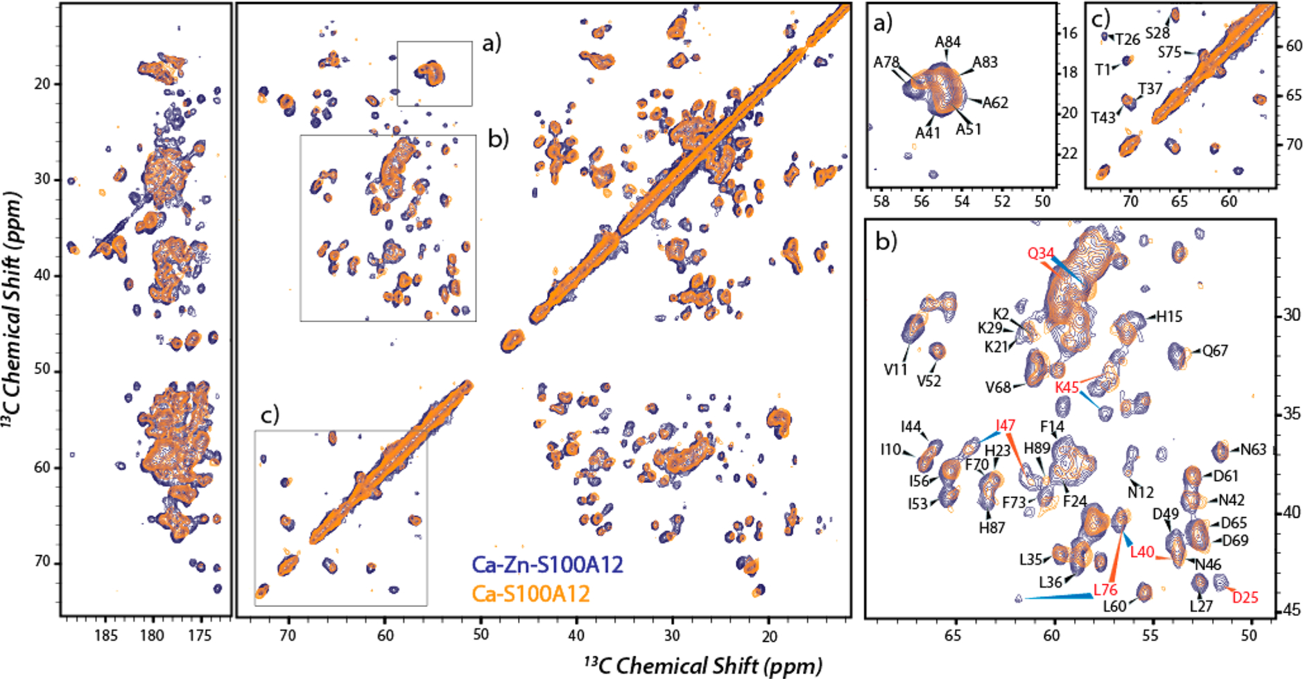
Overlay of 13C–13C correlation MAS NMR spectra of Ca2+,Zn2+-S100A12 (blue) and Ca2+-S100A12 (orange). Panels a–c are close-ups with assignments showing perturbed residues. Residues marked in red show significant CSP upon zinc binding. The experimental conditions were the same as those described for Figure 5.