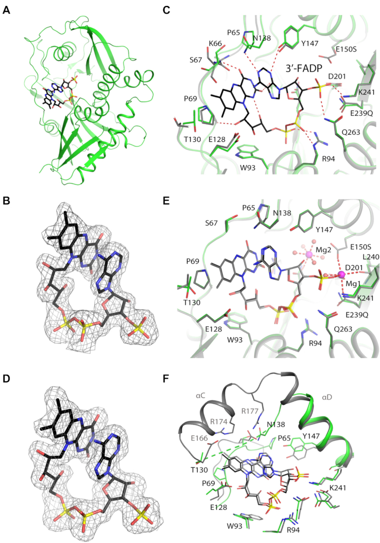
Figure 3.

Crystal structures of SpRai1 in complex with 3′-FADP. (A) Overall structure of SpRai1 E150S/E199Q/E239Q mutant (green) in complex with 3′-FADP (in black for carbon atoms). (B) Simulated annealing omit Fo– Fc electron density at 1.9 Å resolution for 3′-FADP in the complex with mutant SpRai1, contoured at 2.5σ. (C) Overlay of the active site region of SpRai1 in complex with 3′-FADP (in color) with that of free SpRai1 (gray; PDB: 3FQG) (20). (D) Simulated annealing omit Fo– Fc electron density at 1.9 Å resolution for 3′-FADP in the complex with wild-type SpRai1, contoured at 2.5σ. (E) Overlay of the structure of mutant SpRai1 in complex with 3′-FADP (in color) with that of wild-type SpRai1 in complex with 3′-FADP (gray). The magnesium ions from the wild-type SpRai1 structure are shown in pink. (F) Overlay of the active site region of SpRai1 in complex with 3′-FADP (in color) with that of mDXO in complex with 3′-FADP (gray).
