Figure 2.
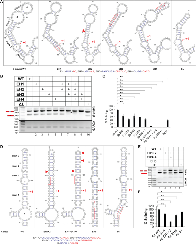
Secondary structure models and splicing activities of pre-mRNAs. (A) SHAPE-derived secondary-structure model of the WT β-globin segment immediately upstream of the 5′-SS (leftmost panel); the designation of loops and stems is noted; anticipated secondary structure models (based on the WT structure) of the exonic segment upstream of the 5′-SS of the mutants corresponding to hybridization/deletion of loops (EH/ΔL) are shown (remaining panels); substituted and deleted native sequence in the mutants are indicated with red rectangle and red triangle, respectively; the original and mutated sequences are shown at the bottom (also see Supplementary Figure S2A); the first nucleotide of the intron is marked with ‘+1’. (B) A representative image showing splicing products of WT, EH and ΔL mutants of β-globin in transfection-based splicing assay; asterisks indicate aberrantly processed products; pre-mRNA and mRNA positions are marked with a red schematic; GAPDH amplification performed as an internal control shown at bottom. (C) Bar-graph showing changes in spliced mRNA products for mutants as a percentage of wild-type RNA and normalized with total RNA generated from the transfected plasmids (n = 3); error bars indicate standard deviation; statistical significance was tested to validate if EH/ΔL mutants of β-globin have lower splicing competence than the WT substrate; ‘*’0.005 < P≤ 0.05, ‘**’P≤ 0.005, N.S. = not significant; the background subtracted gel-images of two additional biological replicates are shown in Supplementary File 8. (D) SHAPE-derived secondary-structure model of WT AdML exonic segment immediately upstream of the 5′-SS (leftmost panel); anticipated secondary structure models of the exonic and intronic hybridization mutants (EH and IH) are shown; original and mutated sequences are shown at the bottom; also see Supplementary Figure S2B. (E) A representative image of splicing products of WT, EH and IH mutants of AdML obtained by transfection-based splicing assay; asterisk and red schematic as in (B). (F) Bar-graph showing quantified mRNA along with the statistical significance of the differences; statistical significance was tested to validate if EH/IH mutants of AdML have lower splicing competence than the WT substrate; the background subtracted gel-images of biological replicates are shown in Supplementary File 8.

