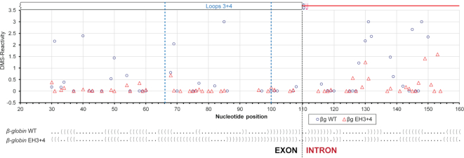
Figure 5.
In vivo structural probing of β-globin WT and its EH3+4 mutant expressed from transfected minigenes. Quantified reverse transcription stops (DMS-reactivity) of individual nucleotides (A & C only) of β-globin WT (o) and EH3+4 mutant (Δ) are plotted against nucleotide positions; dot-bracket notations, which are aligned with the nucleotide positions in the plot, of the SHAPE-derived secondary structure models of protein-free WT β-globin and its EH3+4 mutant are shown at the bottom (dot = unpaired nucleotide, bracket = base-paired nucleotide).

