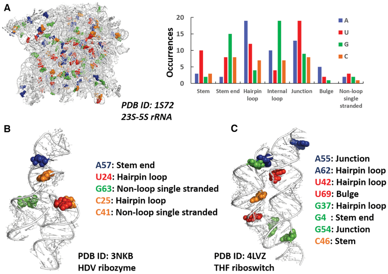
Figure 2.
Water–nucleobase stacking contacts in three case studies. (A) 3D representation of 23S-5S rRNA from H. marismortui (95) with blue, green, red, and orange spheres representing adenine, guanine, uracil, cytosine bases involved in water–base interactions, respectively (left), and number of occurrences of bases involved in the interactions in different structural motifs (right). (B, C) 3D representation of HDV ribozyme (96) and THF riboswitch (97) highlighting the bases involved in water–base stacking contacts, the same bases are listed along with the structural motifs they are located in.

