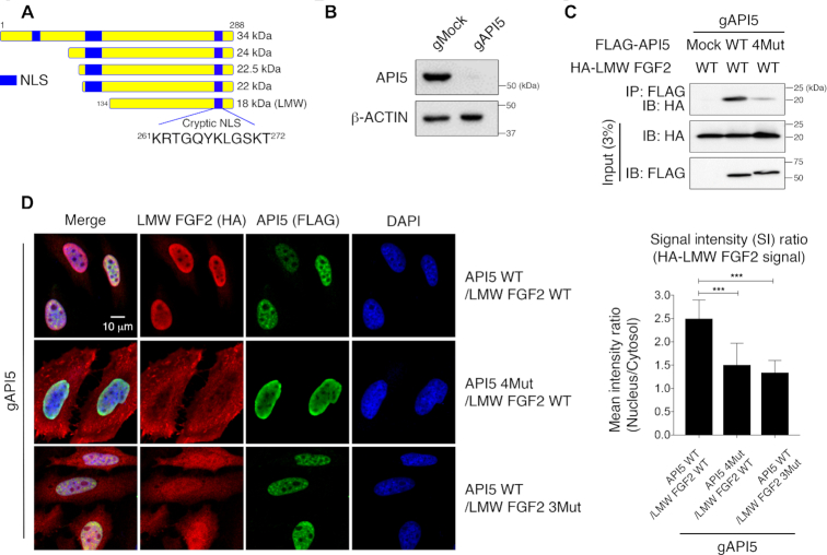
Figure 3.
Cellular localization of LMW FGF2 by API5. (A) Schematic representation of five human FGF2 isoforms and the position of the cryptic NLS region. The cryptic NLS corresponds to the FGF2-segment 1. (B) Western blot validation of HeLa cells after API5 knockout (gAPI5) by the CRISPR/Cas9 system. (C) Monitoring of intracellular interactions of API5 and LMW FGF2 by co-immunoprecipitation assay. Transiently co-expressed FLAG-API5 (WT or 4Mut) and HA-tagged LMW FGF2 WT in API5 knockout (gAPI5) HeLa cells were used. (D) Cellular localization of API5 and LMW FGF2 observed by confocal microscopy and quantified mean signal intensity (SI) ratios of nucleus/cytosol (n = 46 for each sample). The error bars represent the mean ± SD, ***P < 0.001 (Student's t-test).

