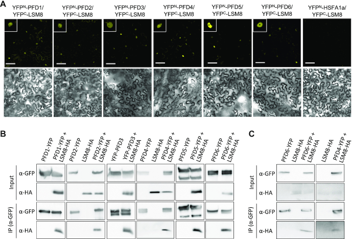
Figure 2.
PFDs interact with LSM8. (A) BiFC assays in N. benthamiana leaves. PFDs fused to YFPN were co-expressed with LSM8 fused to YFPC. Fluorescence from the reconstituted YFP was detected by confocal microscopy from leaf discs three days after infiltration (upper row). YFPN-HSFA1a, which does not interact with LSM8, was used as negative control. Insets show fluorescence from a representative nucleus. Bright field images are shown in the bottom row. Scale bars: 75 μm. (B) Co-immunoprecipitation assays showing the interaction between PFDs fused to YFP and LSM8-HA in N. benthamiana. Total proteins were immunoprecipitated with anti-GFP antibody-coated paramagnetic beads from extracts of infiltrated N. benthamiana leaves. Proteins were detected with anti-GFP and anti-HA antibodies. (C) Co-immunoprecipitation assays showing the interaction between PFD4 and PFD6 fused to YFP and LSM8-HA in transgenic Arabidopsis. Total proteins were immunoprecipitated with anti-GFP antibody-coated paramagnetic beads from extracts of 7-day-old Arabidopsis seedlings. Proteins were detected with anti-GFP and anti-HA antibodies.

