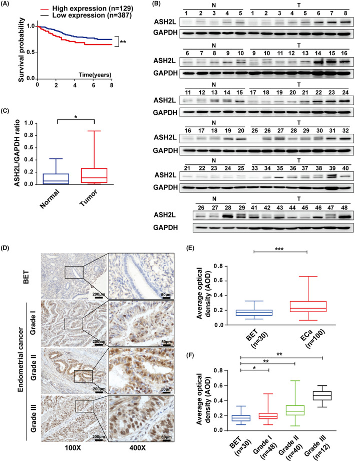
FIGURE 1.

ASH2L is highly expressed in endometrial cancer. A, Kaplan‐Meier analysis for overall survival of 516 endometrial cancer (ECa) patients based on the data from the Cancer Genome Atlas (TCGA). B, Expression of ASH2L in 29 benign endometrial tissues (N) and 48 endometrial cancer (T) detected using western blotting. C, Expression of ASH2L in (B) was quantified using densitometry shown as box and whisker plots. D, Immunohistochemical (IHC) assay in paraffin sections of BET and ECa (histological grades I/II/III) in ×100 magnification (scale bars, 200 μm) and ×400 magnification (scale bars, 50 μm). E and F, Average optical density (AOD) of IHC stained by ASH2L antibody in BET and ECa specimens. Data are shown as mean ± SD, *P < .05, **P < .01, ***P < .001
