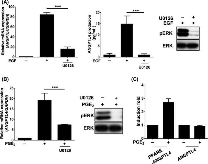
FIGURE 4.

Epidermal growth factor (EGF)‐ and prostaglandin E2 (PGE2)‐induced angiopoietin‐like 4 (ANGPTL4) expression occurs through the ERK pathway. A, B, Cells were treated with 10 μmol/L U0126 and 50 ng/mL EGF (A) or 20 μmol/L PGE2 (B) for 6 h. Expression of ANGPTL4 mRNA and protein was analyzed by real‐time quantitative PCR and ELISA, respectively. Activation of ERK in cells treated with 50 ng/mL EGF or 20 μmol/L PGE2 for 1 h was analyzed by western blotting using anti‐phospho‐(p)ERK1/2 (Thr202/Tyr204) Abs. C, Cells were transfected with 0.5 μg of PGL3 vector containing the ANGPTL4 promoter with an intronic PPAR element (PPARE) by lipofection and then treated with 20 μmol/L PGE2 for 12 h. Luciferase activities were determined and normalized. Values represent mean ± SEM of 3 replicate analyses. ***P < .001
