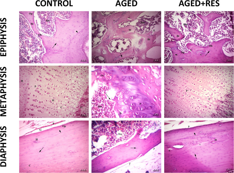
Fig. 2.
Histopathological photomicrographs of the studied groups. The upper left panel: the epiphysis of the control group showing the bone marrow spaces (Bm) between the irregular branching and anastomosing bone trabeculae (Bt) of cancellous bone. Osteoprogenitor cells (Op) and osteoblast (Ob) lining the endosteum with osteocytes inside the lacunae (↑) of bone trabeculae are seen. Cement lines are also seen (arrow head). Notice that the bone marrow is formed of hematopoietic tissue, scattered adipocytes, and blood cells. The upper middle panel: the epiphysis in aged group showing thin bone trabeculae (Bt) surrounding wide fatty bone marrow spaces (Bm). Refractile areas (*) appear inside the trabeculae. Osteoporotic cavities (C) and woven bone (W) in the trabeculae can be seen. Apparent decreased osteocytes (arrow) and eroded areas can also be seen (E). Notice that the bone marrow is fattier as compared with control. The upper right panel: the epiphysis in the resveratrol treated aged rats showing branching bone trabeculae (Bt) enclosing the bone marrow (Bm) spaces. The osteogenic cells (Op) lining the trabeculae are seen. Apparent increase in the number of osteoblasts (Ob) lining the endosteum of the bone trabeculae and the number of osteocytes (arrow) as compared to aged group. Cement line are present (arrow head). Small area of refractile bone (*) and few osteoporotic cavity (C) are still seen. The middle left panel: the metaphysis (epiphyseal plate) in control group showing the resting zone (R), the proliferating zone (P), the hypertrophic zone (H), and the calcified zone (C), followed by the zone of ossification(O). The regularly arranged cell columns with a basophilic matrix are detected. The middle central panel: the metaphysis in aged group showing irregularly arranged columns of cells in the proliferating zone (P) with degenerated cells (D). Wide bone marrow (Bm) and wide empty lacunae (*) are seen. Tear in bone (→) and osteoclasts (Oc) are seen. The middle right panel: the metaphysic in resveratrol treated group showing more regularity of both cells and columns in the proliferating (P) and calcification (C) zones compared with aged groups. The lower left panel: the diaphysis (shaft of femur) in control rat showing outer periosteum (P), Subperiosteal groove (G) and Haversian systems (H). Osteocytes in their lacunae (arrow), osteoprogenitor (Op) and regularly arranged collagen fibres (C) are seen. Cement lines (arrowhead) are detected. The lower middle panel: the diaphysis in aged rat showing eroded periosteum (P) and thinning of the outer fibrous layer(f) of the periosteum. An apparent decrease in the number of irregularly arranged osteocytes (↑) and fattier bone marrow (Bm) as compared with that of the control group . Notice that the shaft is apparently thinner than control. The lower left panel: the diaphysis in resveratrol treated aged rat showing outer periosteum (P) and many Haversion system (H). Nearly normal osteocytes in their lacunae (arrows) with an apparent increase in the number of osteocytes compared with that of the aged group and distinct cement line (arrow head) can be seen. Small osteoporotic cavities (C) and small area of osteolysis that appears as a palely stained area (*) are still detcted (H&E 200X)

