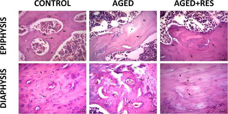
Fig. 3.
Anti-osteoporotic effect of resveratrol in aged rat femur in histological photomicrographs. Upper left panel: the epiphysis in the control group showing the Bone marrow spaces (Bm), irregular branching and anastomosing bone trabeculae (Bt). Osteoprogenitor cells (Op) and osteoblast (Ob), osteocytes (↑) and cement lines (arrow head) are also seen. Upper middle panel: the epiphysis in aged group showing thin bone trabeculae (Bt) surrounding wide fatty bone marrow spaces (Bm). Refractile areas (*), osteoporotic cavities (C), apparent decreased osteocytes (arrow) and eroded areas(E) can also be seen. Notice the few osteoprogenitor cells (Op) at endosteum. Upper right panel: the epiphysis in the resveratrol treated aged rats showing branching bone trabeculae (Bt) enclosing the bone marrow (Bm) spaces. The osteogenic cells (Op), apparent increase in the number of osteoblasts (Ob),the osteocytes (arrow) and cement line are present (arrow head). Small few osteoporotic cavities (C) can still be seen. Lower left panel: the diaphysis of a control rat, showing osteocytes (↑) around a centrally located Haversian canal (H). Cement lines (arrow head) and collagen fibres (L) are noticed. Lower middle panel: the shaft of a rat of aged group showing the eroded periosteum (P) with few osteoprogenitor (Op), osteoblast (Ob), multinucleated acidophilic osteoclast (Oc) in Howships lacunae and bone marrow (Bm. Less acidophilic bone matrix (I)) and multiple osteoporotic cavities containing osteoclast (C) can still be seen. Lower right panel: the shaft of a rat of resveratrol treated group showing many Haversion systems (H). Nearly normal osteocytes in their lacunae (arrows) with an apparent increase in their number as compared with that of the aged group and distinct cement line (arrow head) can be seen. Small area of osteolysis that appears as a palely stained area (*) is detected. (H&E 400X)

