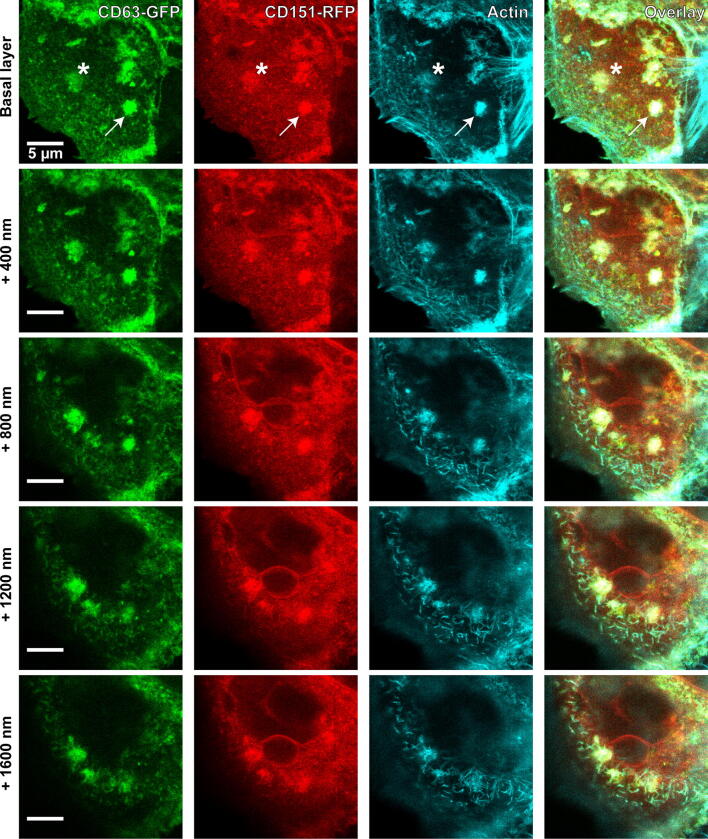
Fig. 3.
Aggregates enriched in CD63, CD151 and actin. HaCaT cells were transfected with CD63-GFP and CD151-RFP, and 1 day later incubated for 3 h at 37 °C with PsVs. Cells were fixed and stained for actin by fluorescently labelled phalloidin, and GFP- and RFP-signal was enhanced by nanobodies. An image stack was recorded starting at the basal cell membrane, with 400 nm axial distances between the optical sections. The linear lookup tables illustrate the channels for CD63-GFP, CD151-RFP and actin in green, red and cyan, respectively. Overlap between all three channels is illustrated in white. See Fig. 4 for the analysis of the relationship between the signals

