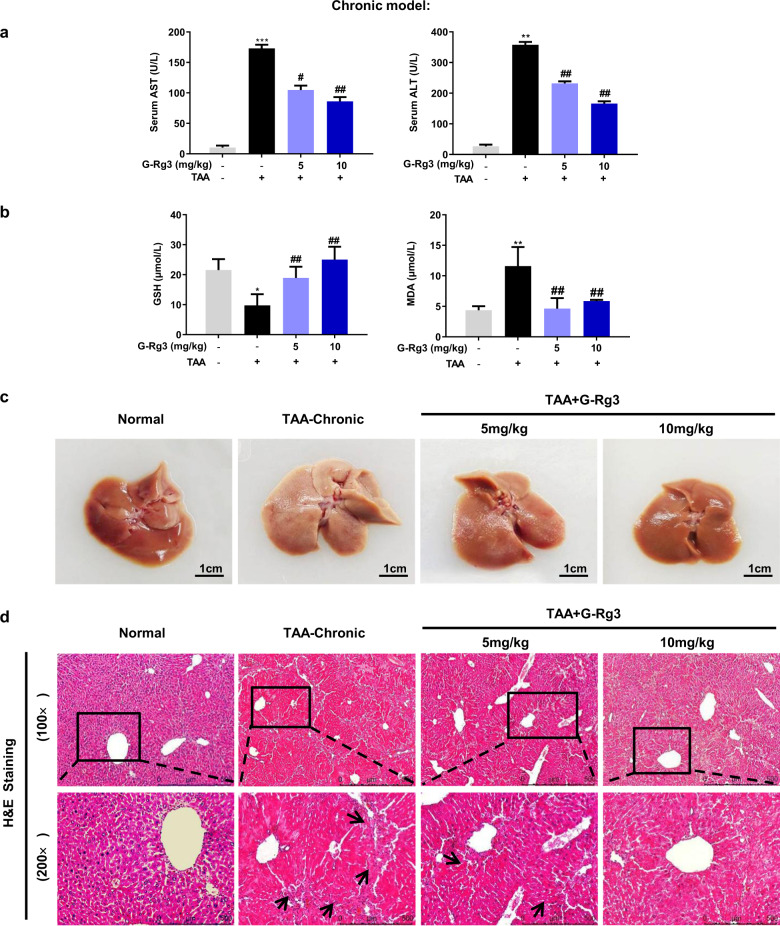
Fig. 2. G-Rg3 ameliorated TAA-induced chronic hepatic injury in mice.
Serological and histopathological examinations were performed with appropriate commercial kits. a Serum level of AST and ALT were tested by biochemical kit. b Oxidative stress levels were tested for GSH and MDA in mice. c The difference in macroscopic pathological observation of liver in groups of Normal, TAA-chronic model, TAA + G-Rg3 (5 mg/kg) and TAA + G-Rg3 (10 mg/kg) were shown after sacrifice. d Histopathological evaluation for liver damage. Representative images of liver sections of different groups stained with H&E staining with amplification of ×100 and ×200. Black arrows indicated long fiber interval. Values were shown as the mean ± S.D (n ≥ 8); *p < 0.05, **p < 0.01, ***p < 0.01 versus Normal group; #p < 0.05, ##p < 0.01, versus TAA-chronic group.

