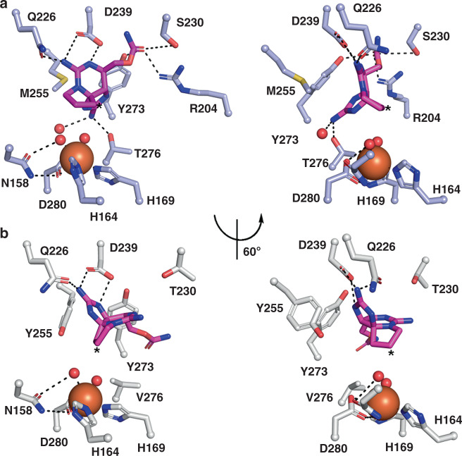
Fig. 4. Structures of SxtT and GxtA with ddSTX bound reveal the protein interactions that are important for correctly positioning substrate for activation.
a ddSTX (dark pink sticks) is anchored in the active site of SxtT via interaction with several different residues that are highlighted here. A rotated view of the active site reveals that C12 (asterisk) is the closest position of ddSTX to the non-heme iron site. b The positioning of ddSTX in the GxtA active site places C11 (asterisk) closest to the iron center. Relative to the placement of ddSTX in SxtT, the guanidinium group that interacts with Thr276 rotates nearly 120º to istead interact with Gln226 and Asp239 in the GxtA active site. Both of these residues are conserved in SxtT.

