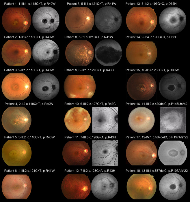
Figure 2.
Fundus photographs and fundus autofluorescence images from 18 patients with CRX-associated retinal disorder (CRX-RD). Fundus photographs and fundus autofluorescence (FAF) images demonstrate macular atrophy in nine subjects (Patients 1, 3–6, 8, 15, 17, 18) and slight atrophic changes at the macula in three subjects (Patients 7, 11, 12). Peripheral atrophy is observed in four subjects (Patients 5, 13, 14, 16; detected by fundoscopy in Patients 5 and 14). Atrophic changes affecting the entire retina, including the macula, mid-periphery, and periphery are found in Patient 5. Macular atrophy is more evident on FAF images in eight subjects (Patients 1, 3, 8, 11, 12, 15, 17, 18). A ring of high density AF is observed in 11 subjects to various degrees (Patients 1, 2, 3, 7, 8, 11–13, 15, 17, 18). Foveal appearance is relatively preserved in nine subjects (Patients 1–3, 7, 10–12, 16, 17).

