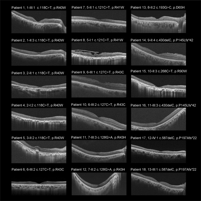
Figure 3.
Spectral-domain optical coherence tomographic images from 18 patients with CRX-RD. Spectral-domain optical coherence tomographic images demonstrate outer retinal disruption at the macula in eight subjects (Patients 1, 3, 4–6, 8, 15, 18). Outer retinal disruption at the peri-macula is observed in 12 subjects (Patients 1–6, 8, 13–15, 17, 18). Intraretinal micro-cystic changes are noted in Patient 13. Epiretinal membrane is found in Patient 8. Marked preservation of the photoreceptor ellipsoid zone (EZ) line at the fovea is identified in eight subjects (Patients 2, 5, 7, 10–14), and slightly preserved EZ at the fovea isobserved in three subjects (Patients 9, 16, 17). Preserved foveal structure surrounded by parafoveal atrophy (i.e., bull’s eye pattern) is found in six subjects (Patients 1, 2, 10, 11, 12, 17).

